


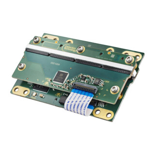
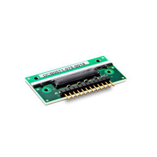
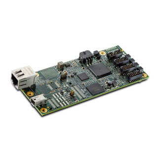
X-Panel 4386a FPM-CSV-TG-S
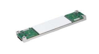
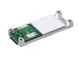
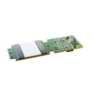
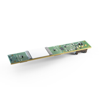
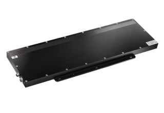
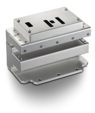
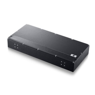
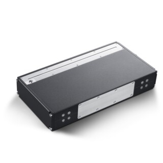
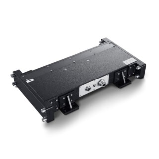
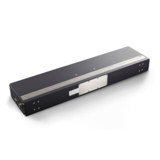
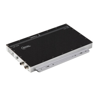
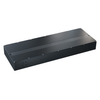
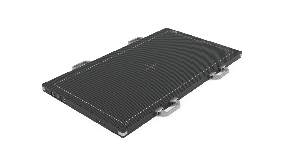
X-Panel 4386a FPM-CSV-TG-S is a very large dynamic amorphous silicon (single-glass a-Si-TFT) flat panel detector (FPD) with direct-deposition CsI scintillator optimized for kV CBCT imaging in radiotherapy.
| Product | X-Panel 4386a FPM-CSV-TG-S |
| Technology | |
| Sensor | Single-glass a-Si-TFT Panel |
| Scintillator | CsI |
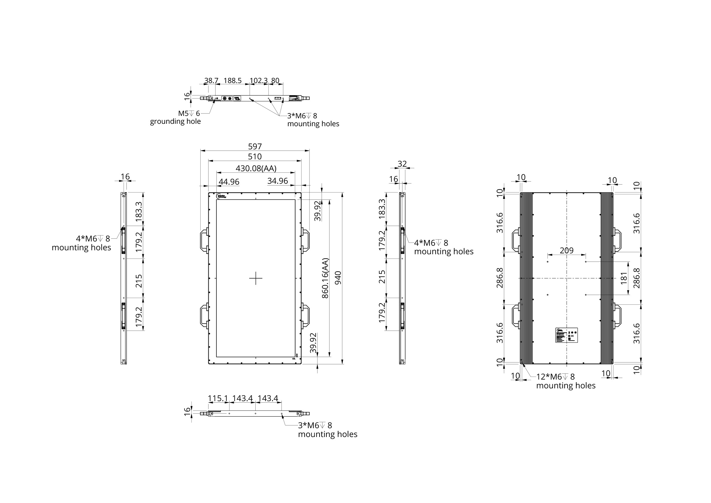
| Active area | 430 x 860 mm |
| Pixel matrix | 3072 x 6144 |
| Pixel pitch | 140 μm |
| ADC | 16 bits |
| Interface | |
| Data interface | 10 G Ethernet |
| Max frame rate | 15 fps (1×1) / 30 fps (2×2) / 45 fps (3×3) / 60 fps (4×4) Higher frame rates with ROI modes |
| Mechanics | |
| Dimensions (H x W x D) | 940 x 510 x 32 mm |
| Weight | 52 kg |
| Environmental | |
| Temperature | 10~40°C (operating); -20~50°C (storage) |
| Humidity | 30~75% RH (operating/non-condensing) |
| IP rating | IPX0 |
| Power | |
| Consumption | 42 W |
| Applications | kV CBCT imaging in IGRT |
FAQs
1. What is the X-Panel 4386a FPM-CSV-TG-S?
X-Panel 4386a FPM-CSV-TG-S is a dynamic, extra-large flat panel detector for kV cone-beam CT (CBCT) imaging in radiotherapy. It shares the single-glass a-Si TFT sensor, CsI scintillator and 430 × 860 mm active area of the X-Panel 4386a FPM-CSV-TG. The “-S” variant adds internal shielding that protects the detector electronics in the radiation environment of a treatment room.
2. Why does the detector have internal shielding?
On a linear accelerator (linac), the kV imaging detector is exposed to radiation scattered from the MV treatment beam. Over time, this scatter can damage or degrade unprotected readout electronics. The internal shielding protects the electronics from MV scatter, which helps maintain stable image quality and supports a long service life in the treatment environment.
3. Which radiotherapy applications is it designed for?
It is designed for kV CBCT in image-guided radiotherapy (IGRT), including patient positioning before treatment and imaging for adaptive radiotherapy workflows. It suits OEMs developing new linac imaging systems as well as those upgrading from 43 × 43 cm kV imagers.
4. How does a 43 × 86 cm detector benefit radiotherapy imaging?
A wider detector captures more of the patient’s cross-section in every view. Depending on system geometry, this extends the reconstructed field of view from about 25 cm with a typical 43 × 43 cm detector to up to 50 cm. Imaging the full cross-section of large patients and anatomies such as the pelvis and abdomen can reduce truncation artifacts, which matters when CBCT images are used for adaptive planning and dose calculation.
5. What frame rates does it support?
The detector reaches up to 15 fps at full resolution, 45 fps with 3×3 binning and 90 fps with 6×6 binning. Region of interest (ROI) modes offer further flexibility in balancing field of view and speed. Higher frame rates allow more views per rotation, which can improve angular sampling and image quality. On faster-rotating gantries, they can also shorten the imaging step of each treatment session and reduce motion artifacts.
6. Why does single-glass TFT matter for radiotherapy CBCT?
The sensor is made from one continuous TFT glass rather than several tiles joined together. That means there are no seams or gaps in the image, which in tiled detectors can cause artifacts in reconstructed volumes. A uniform response across the active area also gives a consistent starting point for calibration. CT number accuracy in the final images also depends on scatter, beam hardening and the system’s reconstruction and correction methods. The detector’s built-in anti-scatter grid mounting helps address scatter.
7. Why a CsI scintillator?
Columnar CsI channels light toward the pixels with little lateral spread. This gives high spatial resolution and high X-ray absorption efficiency at kV imaging energies. It supports good image quality at the low imaging doses used in daily IGRT.
8. Does the detector support an anti-scatter grid?
Yes. The detector has built-in mounting points for an anti-scatter grid (ASG), so OEMs can integrate a grid without designing a custom mounting. Scatter reduction is especially valuable in large-field-of-view CBCT, where scatter from the patient lowers contrast and CT number accuracy. If needed, Detection Technology can connect OEMs with partners who supply large-format grids optimized for the application.
9. How does it integrate into a linac imaging system?
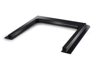
The detector uses a 10G Ethernet data interface and comes with Windows and Linux SDK support. It can be installed in portrait or landscape orientation; in CBCT systems it is typically oriented to maximize the axial field of view. Removable handles make installation on the gantry imaging arm and field service easier. Detection Technology’s application engineers can support OEMs with detector evaluation and integration.
10. What are the detector's dimensions and weight?
The detector measures 940 x 510 x 32 mm and weighs 52 kg. OEMs can take these figures into account when designing the gantry imaging arm and its positioning mechanics.
11. What regulatory standards does the detector meet?
The X-Panel 4386a FPM-CSV-TG-S is a component for medical imaging systems, designed to comply with IEC 60601-1 and IEC 60601-1-2, as well as RoHS, REACH and WEEE requirements. The system manufacturer remains responsible for the regulatory clearance of the final medical device. Detection Technology provides the documentation needed to support the OEM’s submission.