

Western Blot imaging reimagined: How flat panel detectors are transforming protein analysis
Written by Milja Kurkela, Application Engineer at Detection Technology | 16 December 2025

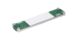
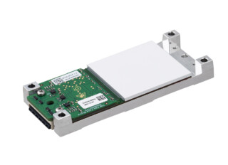
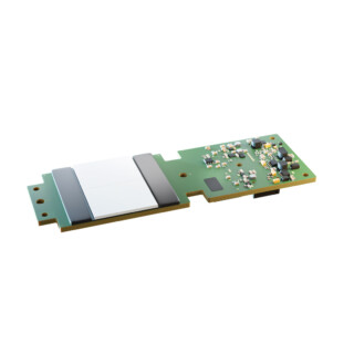
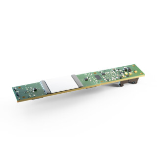
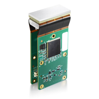
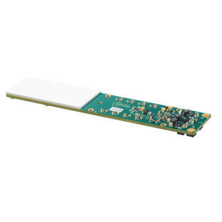
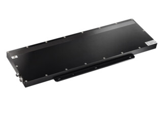
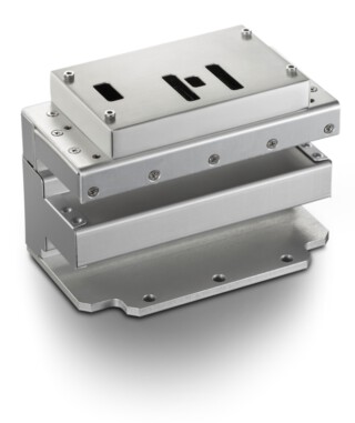
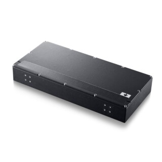
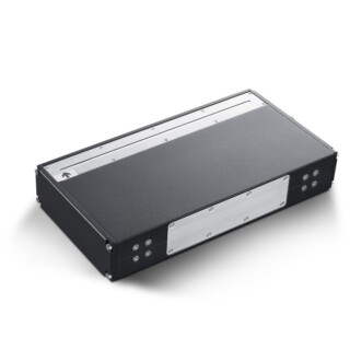
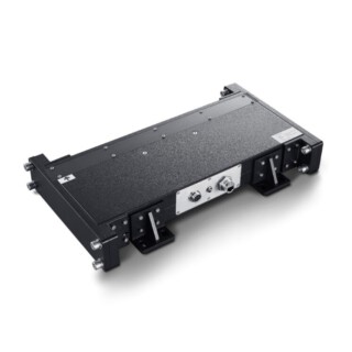
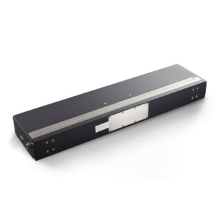
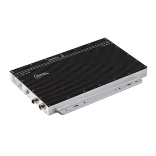
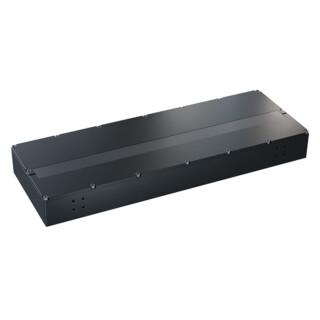
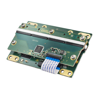
Western blotting remains a cornerstone in molecular biology, but the way we visualise and quantify proteins is evolving rapidly. Gone are the days of darkrooms and film—today’s laboratories demand speed, accuracy, and reproducibility. Enter the X-Panel 1412c FDW: a flat panel detector designed to bring Western blot imaging into the digital age.
Why modernise Western Blot imaging?
Traditional film-based imaging, once the gold standard, now struggles to keep up with the needs of high-throughput labs. Film is slow, has a limited dynamic range, and introduces variability between runs. Even CCD cameras, while an improvement, can fall short in sensitivity and workflow efficiency.
Digital imaging with flat panel detectors changes the game. The X-Panel 1412c FDW, for example, eliminates the need for film exposure and darkroom development. Instead, it delivers immediate, quantifiable results—helping researchers move from experiment to insight faster than ever.

Streamlining the workflow
The Western blot process is familiar: sample preparation, gel electrophoresis, protein transfer, antibody probing, and detection. What’s new is how the signal is captured. With the X-Panel 1412c FDW, the membrane is placed directly onto the detector’s surface. The chemiluminescent signal is captured in real time, producing a high-resolution digital image without extra optical hardware or manual steps.
This direct-to-digital approach means:
- No film or darkroom required
- Instant results for analysis and sharing
- Consistent, reproducible images every time
Real-world validation: Collaboration with the University of Oulu
To put the X-Panel 1412c FDW to the test, Detection Technology partnered with the University of Oulu. The goal: determine if a fully digital workflow could match or exceed the sensitivity, dynamic range, and reproducibility of traditional methods.
The results were clear. The detector captured both strong and faint protein bands with high sensitivity—even low-abundance targets like HIF-1alpha and phosphorylated STAT3 were easily visualised. Integration times as short as one second were sufficient, and the wide dynamic range allowed for simultaneous detection of strong and weak signals in a single exposure.


Performance that speaks for itself
- High sensitivity: Detects low-abundance proteins with clarity
- Wide dynamic range: Captures both strong and weak signals in one shot
- Efficiency: Reduces hands-on time and delivers instant digital results
- Reproducibility: Minimises variability between runs
Labs adopting this technology can expect faster turnaround, improved data quality, and a streamlined workflow—making it an attractive option for both research and diagnostic settings.
Looking ahead
The future is bright for digital Western blot imaging. Ongoing research aims to expand compatibility with new substrates, integrate automation for high-throughput environments, and enhance quantitative analysis software. Long-term stability studies and cross-platform validation will further strengthen confidence in this approach.
Ready to leave the darkroom behind? Discover how the X-Panel 1412c FDW can transform your Western blot workflow and help you achieve faster, more reliable results.